|
|
Current
Research Interests :
|
|
|
Ongoing Projects : |
|
|
Head Pose Estimation and
Face Tracking
Hysteroscopy Video Summarization and Analysis
Intelligent Transportation Systems: Vehicle Tracking and Analysis of Vehicle
Behavior
Object Oriented
Video Coding and Representation
Watershed Drusen
Detection In Eye Fundus Images
Identification of Retinal Structures and Lesions in Color Eye Fundus Images
Color Image Segmentation
Medical
Image Denoising and Enhancement
Pigmented Skin Lesions Classification using Standard Color Camera Images
Visual
Information Retrieval
Medical
Visual Information Exchange on the WEB
|
Head
Pose Estimation and Face Tracking
PROJECT DESCRIPTION:
In this project we investigate
new methods to compute the head pose in monocular images by comparing
the positions of specific facial features with the positions of these
facial features in multiple instances of a prior 3D face model. Given
an image containing a face, we locate facial features such as nose,
eyes, and mouth. Then these 2D feature locations are used as
references in the comparison with the corresponding feature locations
in multiple instances of our 3D face model, projected on the 2D image
space. To estimate the depth of these feature points, we use the 3D
spatial constraints imposed by our face model (e.g. eyes are at a
certain depth with respect to the nose, and so on). The head pose is
estimated by minimizing the comparison error between the face feature
locations in the image and in a given instance of the face model. Our
preliminary experimental results are encouraging, and suggest that our
approach potentially can provide accurate results.
|

|
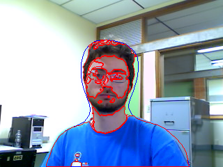
Some preliminary results are illustrated above: (a),(d) and
(g): Original face images; (b), (e) and (h): Best matching
mask instances; and (c),(f) and (i): Best matching mask
instances overlayed on the faces. |
|
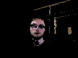
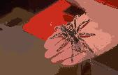
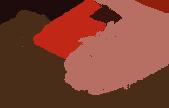
We intend to evolve this work and track faces in different poses in
video sequences. At this stage of our work we are investigating face
segmentation methods based on the detection of skin regions in
monocular images. Below, some
preliminary results
of a segmented head and torso are shown:
(left)
original video frame ;
(center)
detected skin regions ;
(right)
segmented
head.
|
|
|
|
PROJECT DESCRIPTION:
Hysteroscopy is a surgical procedure in which a gynecologist uses a small
lighted telescopic instrument called a hysteroscope
to diagnose and treat many uterine disorders. A hysteroscopic
examination has different phases, and usually only the relevant findings are
used for the diagnosis and prognosis in fertilization studies and in the
Gynecology practice. Consequently, most visual information in such videos is
not relevant, and only a reduced number of frames are used. Currently, a
summary of a hysteroscopic video is obtained manually during the diagnosis
process, by selecting only the relevant frames of the different phases of a hysteroscopic procedure, and later the findings in these
frames are reported and described in the patient records. Therefore, in this
project we intend to develop a simple and accurate method to extract concise
representations of hysteroscopic videos contents
that could be used in the clinical practice.
Application:
Given a library with thousands of diagnostic hysteroscopy videos, which are
only indexed according to a patient ID and the exam date. Usually, users
browse through this library to obtain answers to queries and retrieve images
of submucosal myomas or recover images whose diagnosis is endometrial polyp.
This work will allow to identify clinically relevant information in diagnostic
hysteroscopy videos, since only portions of the recorded videos are relevant
from the diagnosis/prognosis point of view. Our goal is to capture the
specialist intention by tracking image points through the image sequence. We
demonstrate that the resulting representation is a helpful way to organize the
hysteroscopy video content, allowing specialists to perform fast browsing
without introducing spurious information in the video summary. The preliminary
experimental results indicate that our method produces compact video summaries
(data-rate reduction around 97.5%) without discarding clinically relevant
information.
PRELIMINARY RESULTS:
|
Uninteresting frame
|
Interesting
frames provided by our method
|
|

|

|

|
|

Video
segment trees computed for a particular hysteroscopy video. X-axis
represents the frames in the video sequence. Red line segments represent the
video segments manually selected by specialists. Notice the relevant video
segments appear associated with taller trees. |
|
|
|
Intelligent Transportation Systems: Vehicle Tracking and Analysis of
Vehicle Behavior
PROJECT DESCRIPTION
:
Intelligent transportation systems (ITS) are
systems that use advanced computer vision techniques to gather
traffic monitoring
information. These information can be used in surveillance, traffic
control and identification of unusual vehicle behavior, among others
applications. Vehicles tracking is a fundamental task in
ITS, because it allows to identify the vehicle
location at any time. However, the vehicle tracking process can be
interrupted under different types of occlusions, like bridges, traffic
signs and other vehicles on the road. We are developing a new adaptive
particle filter approach to handle vehicle occlusions, and we also are
working on the analysis of vehicle behavior using the obtained vehicle
tracking information with promising results.
PRELIMINARY
RESULTS:
|
|

Tracking results for a
video sequence containing vehicle occlusions.
On the left, there are vehicle tracking results
under several occlusions (frames 03 (a), 35 (b) and 55 (c) of the video
sequence). Our Adaptive Particle Filter approach is
able to resume the vehicle tracking process after the the vehicle becomes
disoccluded. The solid/dashed line shows the vehicle tracking results
obtained by our adaptive particle filter.
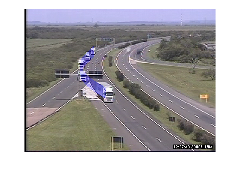

Analysis of vehicle behavior. On the top a
truck changes lanes
dangerously (blue ribbon). Below it, a truck is tracked even under the
occlusion by a bridge (green ribbon). These figures show the vehicles path
and a “stroboscopic effect” illustrating the vehicle motion and behavior.
|
|
Object Oriented
Video Coding and Representation
PROJECT DESCRIPTION:
Object video representation provides a
convenient approach for several compression and transmission tasks. However,
the coding efficiency is directly related to the effectiveness of the object
segmentation. In this work, we develop a video coding approach using video
segmentation based on motion coherence. Long-range motion patterns are
identified by computing correspondence of sparse points (i.e. particles) and
the segmentation of particle trajectories using an ensemble clustering
approach. Then, a dense video frame representation (i.e. a pixel-wise
representation) is obtained, leading to object tunnels in the spatio-temporal
domain (see figure below). Instead of motion boundaries, the segmentation is
guided by the consistent motion behavior of sample points of the frames. This
strategy allows to extract longer tunnels in the spatio-temporal domain. The
proposed approach generates a simple scene representation, adequate for object
video coding, and also delivers a more redundant and temporally persistent
partition of the scene than direct video segmentation methods and motion
prediction strategies.
PRELIMINARY RESULTS:
|
 |
 |
   |
|
Particles
|
Particle trajectories segmentation
|
Pixel-wise segmentation and object
tunnels 1 and 2
(green and
blue bands, respectively) |
|
|
Watershed Drusen
Detection In Eye Fundus Images
PROJECT DESCRIPTION:
Age-related macular degeneration (ARMD) can evolve rapidly, and cause severe
losses to the central vision of patients. An early sign of ARMD is the formation
of drusen in the retina (i.e. white-yellow spots
located in
the center of the macula). Therefore,
the early identification of drusen in the ARMD
process, and their delimitation and quantification over time, is very important for its medical treatment. This project
is focused on the development of robust methods for drusen detection and segmentation,
delimiting precisely individual
drusen, even when the drusen
spots are spatially close. We intend to improve drusen
segmentation and overcome common drusen segmentation
difficulties reported in the literature.
PRELIMINARY RESULTS:
|
Original Image
|
Literature
|
Our result
|
|

|

|

|
|
| |
Identification of Retinal Structures and Lesions in Color Eye Fundus Images :
Diabetic Macular Edema
PROJECT DESCRIPTION:
In this
project, we are developing automatic methods to solve several problems
related to the detection and classification of the Diabetic Macular Edema
(DME) using color eye fundus images. We have developed
methods to detect the optic disk rim, the fovea center, and
the exudate lesions. Our experimental results are encouraging, and
indicate that our approach potentially can achieve a better performance
than other known methods available in the literature. Some results are
illustrated in Figures 1(a) and 1(b).
PRELIMINARY
RESULTS:
|
 |
|
Figure 1: A typical
eye fundus image. (a) Detected optic disk rim superimposed on the
original color eye fundus image. (b) Exudate lesions and polar fundus
coordinates (which are centered on the fovea center) superimposed on a
color eye fundus images.
|
|
Color Image Segmentation
PROJECT DESCRIPTION:
Image segmentation is, by definition, the
problem of decomposing images into regions that are semantically uniforma
(e.g. correspond to the visible objects in the scene), and is often the
first important step of an image understanding system. The goal of this
project is to develop image segmentation techniques that suitably
separate the relevant classes of objects in a scene, based on color and
spatial information.
Some preliminary results obtained with
our feature space tesselation
approach and posterior data clustering in
joint spatio-color domain are illustrated below.
PRELIMINARY
RESULTS:
|
Original
Images: |
Segmented by our method: |
Segmented
by Mean Shift:: |
|
|
|
|
|
 |
 |
 |
|
|
PSNR: 23.80
Clusters
(colors): 220
Regions: 2480
|
PSNR: 23.22
Clusters
(colors): 5088
Regions: 5196
|
|
|
|
|
|
PSNR: 23.36
Clusters
(colors): 33
Regions: 936
|
PSNR: 18.91
Clusters
(colors): 49
Regions: 63
|
|
| We
are investigating the effect of shading attenuation in low level color
vision, specially in face detection problems. Some preliminary results
obtained with
our shading attenuation
method and and posterior face segmentation are illustrated below.
PRELIMINARY
RESULTS:
|

|
Face segmentation examples. In the first and second columns
the original images nd their respective segmentation results
are shown. In the third and fourth columns, the images after
using our shading attenuation method, and their respective
segmentation results are shown. |
|
|
|
|
|
|
Medical
Image Denoising and Enhancement
PROJECT DESCRIPTION:
This
project is focused on medical image noise suppression and enhancement.
Different image modalities are studied and suitable denoising and
enhancement methods are investigated. Two examples are illustrated below.
IMAGE DENOISING AND
ENHANCEMENT IN MAMMOGRAPHY:
In this work, wavelet based methods are investigated. At each resolution, coefficients associated with noise are modelled by Gaussian random variables; coefficients associated
with edges are modelled by Generalized Gaussian random variables, and a shrinkage function is assembled based on posterior
probabilities. Given a resolution of analysis, the image denoising process is adaptive
(i.e. do not require troublesome parameter adjustments), and the selection of a gain factor provides the desired detail
enhancement. The enhancement function must be designed to avoid introducing artifacts in the enhancement process, which is
essential in mammographic image analysis. We intend to develop techniques
that are easy to use (by mammographists), and can help detecting microcalcifications and
other suspicious structures in situations where their detection would be difficult
otherwise.
PRELIMINARY
RESULTS:
|
 |
 |
|
Original mammographic image |
Denoised and enhanced version showing a
microcalcification cluster (middle right) |
|
|
DENOISING AND ENHANCEMENT OF
PROSTATE ULTRASOUND
IMAGES:
In this study, we investigate the applicability of a Monte Carlo approach to
despeckling transrectal ultrasound (TRUS) images
of the prostate. The particularities and noise statistics of TRUS images of
the prostate are incorporated into a likelihood-weighted
Monte Carlo estimation scheme. Our preliminary in-silico and in-vivo
experimental results are promising, which was confirmed by a clinical
evaluation of the in-vivo test cases, and indicate that our method
potentially can perform better than other state-of-the-art methods recently
proposed.
This
project has been developed in collaboration with Dr. Alexander Wong (U. of
Toronto, Canada).
PRELIMINARY
RESULTS:
|

Despeckled
log-envelope images produced by state-of-the-art tested methods,
and by the proposed
despeckling method. The image
plate shows transverse transrectal ultrasound images of the
prostate
with left posterior adenocarcinomas confirmed by biopsy. |
|
|
|
|
|
|
Pigmented Skin Lesions Classification using Standard Color Camera Images
PROJECT DESCRIPTION:
Pigmented skin lesions have two forms, benign (called nevi) and malignant (called
melanoma). Since melanoma is one of the most dangerous cancers nowadays,
there is a strong interest in digital image analysis systems to help, and
maybe improve, the physician diagnosis and pre-screening. However, most of
these efforts are based on dermoscopy, a noninvasive tool that improves the
recognition of submacroscopic morphologic structures and vascular patterns,
facilitating the use of a computer system in melanoma diagnosis, but with
significant investments. Oour goal in this project is to develop a system to
segment and classify skin lesions just using standard camera images (a
simple color photograph of the skin lesion). Our preliminary results
indicate a pre-screening accuracy of 98%, without any false negatives.
PRELIMINARY
RESULTS:
 |
|
Examples of pigmented skin lesion segmentation. In the
first and second columns, the original images and their respective
segmentation results by state-of-the-art methods. The third and fourth
columns show the
resulting images after the application of our proposed shading attenuation
method, and the respective segmentation results. |
|
Visual
Information Retrieval
PROJECT DESCRIPTION
:
We
are studying content-based indexing and retrieval of compressed color
images, mostly for image retrieval in the World Wide Web and in medical image repositories. We are interested in
multi-resolution techniques that allow user interaction, and are based on probabilistic texture
models.
PRELIMINARY
RESULTS:
|
|
|
|
|
Medical Visual Information Exchange on the WEB
PROJECT DESCRIPTION :
The web has become such an extensive health information repository in the
world that it is increasingly difficult to search for relevant medical
information. Most medical information available on the web is not peer
reviewed, and is retrieved imprecisely by current web search mechanisms
(i.e. based on keywords). This project aims at developing a metadata model
that allows to describe medical visual information (i.e. medical images)
of different modalities, including their properties, components,
relationships, and authorship. The model uses the web architecture and
supports the international classification of diseases and related health
problems (i.e. ICD-10). An RDF schema derived from this metadata model is
integrated to each medical image, and specifies the semantics of each
property in the image. Thus, relevant information can be extracted
directly from the images, and data integrity is better preserved in the
web.
PRELIMINARY RESULTS:
|
|
|
|
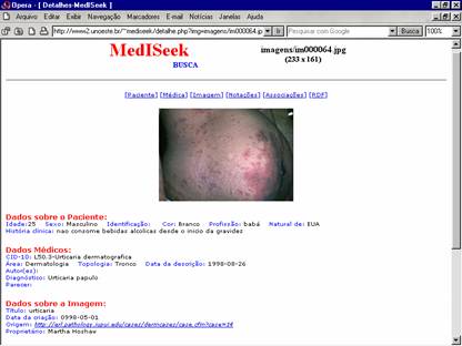
Prototype built (MedISeek)
to validate our metadata model,
and mechanism for medical visual information exchange on the web.
Authorized system users have been able to describe, store and
retrieve medical images and their associated diagnostic information.
|
|